News
EBUS: Device to diagnose diseases without opening up patient’s chest
Not only into the difficult recesses of this organ essential for breathing can this slim wonder-tool delve, but it can also check out adjacent areas to help doctors diagnose diseases without opening up the chest of the patient.
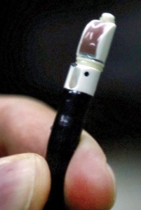
The EBUS scope. Pix by M.A. Pushpa Kumara

Dr Anil Jayasinghe
Now available for the first time in Sri Lanka at the National Hospital in Colombo, is the Endobronchial Ultrasound (EBUS) scope which will take the diagnosis of lung cancer and infections and other diseases causing enlarged lymph nodes in the chest to a different level, the Sunday Times learns.
There is an air of expectancy recently among those gathered at the National Hospital’s third-floor ENT Lecture Hall, while from the Operating Theatre (OT) on the same floor is relayed images of procedures being carried out with the EBUS scope.
In the OT, talking gently to the 33-year-old woman patient is Senior Registrar in Respiratory Medicine, Dr. Yamuna Rajapakse, urging her to concentrate on her breathing. Dr. Rajapakse has just returned from Australia, having been trained in this skill which is relatively new to the world, at the Royal Adelaide Hospital by experts Dr. Peter Robinson and Dr. Phan Nguyen of the Department of Thoracic Medicine.
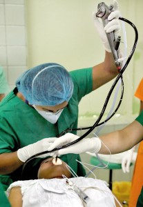
Dr. Yamuna Rajapakse performing the procedure on a patient
The patient’s symptoms have been an intermittent fever and mildly productive cough for about two months and headache. She has not experienced shortness of breath and there has been no known exposure to tuberculosis. She has never smoked and there is no past history of co-morbidities (other diseases). A computed tomography (CT) scan has indicated a mass, posterior to the right main bronchus.
While Dr. Rajapakse tests the 6-mm EBUS scope, the patient is placed under mild sedation. As the scope is gently sent through her mouth, vocal cords, trachea (wind-pipe) and then firstly to the left main bronchus and next to the right main bronchus, close-up views come up on screen.
Dr Amitha Fernando
It is behind the right main bronchus that the mass is spotted through the ultrasound. Thereafter, a needle is passed through the scope to take a few specimens for a biopsy.
At an earlier meeting with the Sunday Times, Senior Consultant Respiratory Physician Dr. Kirthi Gunasekera and Consultant Respiratory Physician Dr. Amitha Fernando talk of the developments in the field of respiratory medicine over the years.
“In the 1950s, there were direct rigid bronchoscopes that doctors worked with to take a look at a person’s lungs,” says Dr. Gunasekera, explaining that the flexible bronchoscopes with fibre-optic cameras came into use in the early 1970s. Till the early 2000s, doctors could look inside the lumen (the space within the tubular structures of the lung) with these.
In a bronchoscopy, when we inserted a rigid or flexible bronchoscope, the area outside the bronchi remained “blind”, stresses Dr. Fernando.Disease diagnosis would be with a CT scan while a biopsy could be taken with this bronchoscope if the suspect area was within the lumen.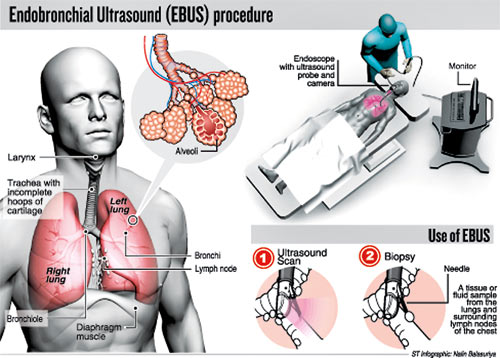
There were limited options and often surgery dubbed ‘mediastinoscopy’ under general anaesthesia had to be performed to get access and examine the inside of the

Dr Yamuna Rajapakse
upper chest between and in front of the lungs (mediastinum). Under this surgical procedure, a small incision would have to be made in the neck just above the breastbone or next to the breastbone and a mediastinoscope put through to see the lungs and surrounding lymph nodes. Tissue or fluid would also have to be collected through a biopsy, it is learnt.
“We used a medical thoracoscope, meanwhile, through a small incision in the chest to look into the pleural cavity and when lesions were found to remove fluid and take biopsies under direct vision,” says Dr. Gunasekera, adding that it was very cost-effective when compared to open surgery.
Now, however, there has been much advancement, reiterates Dr. Rajapakse, and this endoscopic procedure, EBUS, combines the fibre-optic bronchoscopic procedure with ultrasound scanning. (See box)
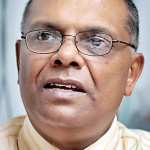
Dr Kirthi Gunasekera
EBUS has been around for about 10 years in centres of excellence such as Japan, the United States of America, the United Kingdom, Australia and closer home in Singapore, the Sunday Times learns.
This advancement in interventional pulmonology, which is a safer outpatient procedure, is available in Sri Lanka and will help numerous patients.
| Advantages of using EBUS The many advantages of EBUS, the first of its kind in Sri Lanka, include the patient not being under general anaesthesia (fully asleep) but under ‘conscious sedation’ where he/she is awake and can respond during the procedure. Life-threatening complications are minimal and the patient spends a shorter time in hospital, says Dr. Yamuna Rajapakse, adding that EBUS is a day-procedure, unlike open surgery after which there would be a stay in the Intensive Care Unit. Explaining the details of the EBUS procedure, she says that they can look into the patient’s lungs as well as take biopsies of the glands in the centre of the chest, lying outside the bronchi (breathing tubes). The EBUS tool consists of a flexible tube (bronchoscope) with a small ultrasound probe at the end of it and a small camera above the ultrasound probe. NHSL should lead in implementing new techniques: Director Reiterating that EBUS is an endoscopy technique combined with the ultrasound technique which allows doctors to go deep into the respiratory system to spot malignancies, National Hospital Director Dr. Anil Jasinghe underscored current thinking that the premier hospital in Sri Lanka catering to patients from all over the country should not lag behind any private hospital. This is why he encourages his Consultants to implement new techniques, so that the National Hospital leads in the provision of health-care. When the NHSL takes the lead, affordability in the private sector becomes standard, he said, adding that “it’s all about technology and skill, while the other aspect is organisation within the NHSL. Although the Consultant Chest Physicians are not attached to the NHSL, they work here simultaneously without any hindrance”. Dr. Jasinghe assured that within about four years he is expecting to establish a chest unit at the NHSL. |

