Thoracoscopy is a novel technique which uses a video camera system to operate inside the chest cavity for a wide variety of lung/chest conditions. The use of camera systems reduces the requirement for large surgical incisions which we use to enter the chest cavity.
In conventional Thoracotomy -the incision used to enter the chest cavity is about 10-15 cm in size and the surgeon has to cut through several groups of muscles and nerves. This can result in complications such as increased pain, scar tissue formation, infection and damage to nerves and muscles, thereby affecting their normal function. This may require a longer stay in hospital and slow recovery.
A smaller incision reduces the stay in hospital and the patient will be able to go home sooner even if the operation is complex. But it must be remembered that this approach needs to take into account the patient’s problem and suitability and may not be possible in all occasions.
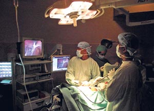
Thoracoscopy set-up
- Especially made small (10mm diameter) camera
- Instruments to manipulate tissues and internal organs
- Video monitoring and recording
- Computer system for image processing and patient data collection
Thoracoscopy uses small 1cm, two or three cuts strategically placed on the chest wall to introduce the camera and instruments into the chest cavity. Small trocars are used to enter the chest cavity through 1cm incisions without damaging the muscles and nerves. Unlike a conventional thoracotomy, the camera can inspect all nooks and corners of the chest cavity giving a magnified view, resulting in better detection and assessment of the disease process.
As it does not require cutting through several groups of muscles , entering the chest cavity is quicker and simple with reduced blood loss (during conventional thoracotomy , blood loss can be 200ml-500ml compared to 2-5ml loss during thoracoscopy). We could say that the blood loss is negligible.
Thoracoscopy will reduce the operating time for a given operation by half (between 1-2 hrs spent on cutting and suturing muscles during conventional thoracotomy).
When the procedure is over, it only requires two to three stitches to close up the skin wound (as the muscles are not cut , it does not require any suturing).
Long term disability due to muscles and nerve damage is non-existent.
Disadvantages of Thorocoscopy
These are very minimal. However, the surgeon should be familiar with the equipment and the internal anatomy of the chest cavity. There is a learning curve for junior surgeons. There is a risk that the procedure may have to be converted to a conventional procedure if there is bleeding or doesn’t provide adequate access.
Types of operations that
can be performed
Almost all the operations inside the chest cavity including removing part of the lung (lobectomy) for lung cancer can be performed using the thoracoscope.
Operations that are performed using the thoracoscope can be done for diagnostic or therapeutic purposes.
Previously almost all diagnostic procedures like biopsies (taking small part of the suspected disease tissues to inspect under microscope to come to a diagnosis) needed a conventional thoracotomy. CT scan guided biopsies are available but not always possible.
In modern day thoracic surgery, performing a thoracotomy for a biopsy is unacceptable. Almost all diagnostic procedures can be carried out using the thoracoscope.
Although most of the therapeutic procedures are done under a general anaesthetic, diagnostic procedures can be carried out under sedation.
Diagnostic procedures
- Removal and testing of fluid collections inside the chest cavity
- Lung biopsies
- Mediastinal (structures in the middle of the chest cavity) Lymph node Biopsy
- Biopsy of Mediastinal Tumours
- Determining the extent of spread of lung cancer
Most of these diagnostic procedures can be done within 30 minutes by a Thoracic Surgeon who is trained in Video Assisted Thoracoscopic Procedures (VATS ).
Depending on the findings during the diagnostic procedure, the Thoracic surgeon can perform the therapeutic procedure in the same sitting. For example when you diagnose a fluid collection inside the chest due to spread of cancer, the surgeon can perform the operation to evacuate the fluid and prevent further fluid collection in the future at the same sitting.
Every case with suspected mediastinal (inside the chest) lymph node enlargement, needs a thoracoscopic biopsy. CT guided biopsy is difficult and is not adequate in suspected Lymphomas (cancer arising from lymph nodes), sarcoidosis and tuberculosis.
Biopsy of the pleura (lining tissues of the inside of chest cavity) is made easier with a thoracoscope as it allows inspection of every corner of the chest cavity with a magnified view. Diagnostic accuracy is very high.
Therapeutic procedures
These can be divided into three categories:
- pleural diseases ( cavity between lung and chest wall)
- lung diseases
- mediastinal diseases ( middle part of the chest cavity)
The commonest conditions of pleural disease are, air in the pleural cavity (Pneumothorax) and fluid in the pleural cavity (Pleural effusion/ empyema).
Air in the pleural cavity (pneumothorax) is quite common in young adults and is due to rupture of small air filled sacs in the lung. It can also occur in the elderly due to various lung conditions. When the air gets collected inside the thoracic cavity, the lung starts to collapse and the person will have difficulty in breathing and chest pain.
The operation for a pneumothorax using the thoracoscope will take 30-40 minutes with two small 1cm incisions on the chest and includes removing the diseased part of the lungs and removing the pleura (pleurectomy). The patient will be able to go home in three days.
Fluid in the pleural cavity can either be simple fluid (pleural effusion) or pus in the pleural cavity (empyema). Most elderly patients with undiagnosed pleural effusions require a thoracoscopic examination to inspect and take biopsies from the pleural cavity for a diagnosis. And at the same time, the surgeon can perform the definitive procedure to prevent future fluid collections in the pleural cavity.
Pleural effusions in the elderly can be due to lung cancer, cancer deposits from the other organs in the body, tuberculosis, lung infections (pneumonia/lung abcess) etc.
Pus in the pleural cavity (empyema) is due to bacterial infection of the fluid in the pleural cavity. Patient will have high fever, difficulty in breathing and chest pain. Initially this requires drainage by insertion of a tube into the chest cavity. Later the patient will require a definitive operation to cure the condition and to prevent complications.
The majority of empyemas can now be treated with thoracoscopic operations. It is calleded Video Assisted Thoracoscopic Decortication ( VATS Decortication). It requires a small 2-4 cm incision on the chest wall and the patient can go home in 3-4 days. Most of the lung resections including removing the part of a lung for lung cancer (VATS Lobectomy) are now carried out using a thoracoscope.
Thoracoscopic operations are routinely used for removing large air filled cavities in the lung called Bulle. It is also very useful in operating on patients with emphysema (Lung Volume Reduction Operation) as these patients cannot withstand a conventional thoracotomy.
Other lung conditions that can be treated with thoracoscopic operations include Bronchogenic cysts, benign lung tumours, lung secondaries ( cancer deposits in the lung from other cancers elsewhere in the body), Bronchiectasis, lung abcesses and congenital conditions. It can also be used in the treatment of gullet tumours, called an oesophagectomy.
Most of the operable Thymic tumours and Mediastinal cyst and mediastinal tumours except for the huge mediastinal tumours are now routinely removed using Thoracoscope at Chest Hospital, Welisara. Thoracoscopy allows us to operate on patients previously thought inoperable due to co-existing disease conditions.
Introduction of Thoracoscopic operations has changed the conventional way of treating lung diseases. It has changed the management guidelines in treating thoracic disease conditions. The availability of this technique has allowed us to make leaps and bounds the treatment of lung conditions. |