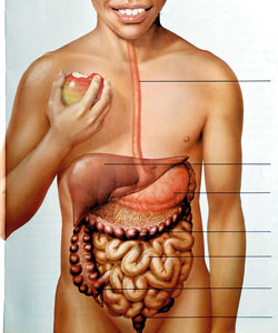
The small bowel (or small intestine), a vital organ involved in nutrient absorption, is the longest portion of the gastrointestinal (GI) tract. Called “small” because it is thin or narrow compared to the “large” bowel (also known as the colon), it is much longer than the large bowel (15 feet on average).
GI bleeding, including from the small bowel, occurs when an abnormality on the inner lining begins to bleed. The bleeding may be slow, resulting in anaemia (a low blood count), or it may be rapid, causing a haemorrhage. Approximately 5% of all GI bleeding comes from the small bowel.
The causes of bleeding in the small bowel are different from those in the colon or the stomach. The most frequent causes of bleeding from the colon are polyps, diverticulosis (small out-pouchings in the wall of the colon), or cancer. Upper GI (esophagus, stomach, or duodenum) bleeding is most frequently due to ulcers.
30 to 40% of small bowel blood loss is caused by abnormal blood vessels that lie within the wall of the small bowel. These abnormal blood vessels are called angioectasias or arteriovenous malformations (AVMs). AVMs become more common as people age and are associated with other medical problems, such as chronic kidney disease and valvular heart disease.
In people over 50, AVMs are the most common cause of small bowel bleeding. Other causes include benign (non-cancerous) and malignant (cancerous) tumors, polyps, Crohn’s disease (a type of inflammatory bowel disease), and ulcers. Like ulcers in the stomach, ulcers in the small bowel are often the result of the use of pain killers called nonsteroidal anti-inflammatory drugs (NSAIDS)
How is the small bowel examined?
There are multiple techniques. In most cases, the first step is endoscopy and/or enteroscopy. If that fails to find the source of bleeding, a common next step is capsule endoscopy. X-ray options include a small bowel follow-through, or a computed tomographic scan (also known as a CT or CAT scan) of the small bowel. Deep small bowel enteroscopy can now be performed using special scopes with inflatable balloons and/or overtubes. The final option, which is usually used only if other methods have failed, is intraoperative enteroscopy.
What are standard endoscopy
and enteroscopy?
Endoscopes and enteroscopes are instruments resembling long, thin tubes with a light and a camera at one end used by doctors to evaluate the stomach and small bowel. Endoscopy refers to the examination of the bowel using a scope. The images obtained are displayed on a monitor. The scopes also have channels which allow instruments to be passed down them to treat lesions, to obtain biopsies, or to mark the location of a lesion with a tattoo to aid a surgeon in locating it.
The examination begins with the patient receiving a sedative. The doctor then passes the scope through the mouth. A regular endoscope is capable of examining the esophagus, stomach and the first portion of the small bowel, known as the duodenum. If the source of bleeding is thought to be lower down in the small bowel, a longer scope, known as an enteroscope, can be used to reach the middle portion of the small bowel, the jejunum.
What types of x-ray studies are
used to find the source
of small bowel bleeding?
X-ray studies continue to have a role in the evaluation of small bowel bleeding, as 20-25% of bleeding that originates in the small bowel is caused by abnormalities in the intestinal wall, such as tumors, that can be seen by specialized x-ray studies. Three x-ray tests are commonly used– small bowel follow-through, enteroclysis, and CT enterography.
The small bowel follow-through test is a series of abdominal x-rays taken at different times after a patient drinks a white, chalky fluid called barium, that shows up clearly on x-rays. The test allows the doctor to examine the lining of the intestine for any irregularities. The test which is safe and easy to tolerate is good for large abnormalities, but can miss many smaller ones.
A second x-ray test, the enteroclysis study also uses barium to visualize the inner wall of the small bowel. It is more invasive because it requires a small tube called a catheter to be slowly advanced from the nose down the esophagus, through the stomach and into the small bowel, to allow for air and barium to be instilled.
The pictures from enteroclysis have better resolution, so abnormalities missed by the small bowel follow-through test may be detected but it can be an uncomfortable examination due to the presence of the catheter and the use of air to distend the small bowel while taking pictures.
A third test, a CT enterography, is done the same way as a normal CT scan. The patient drinks an oral contrast solution (often dilute barium) while also receiving intravenous (IV) contrast. Then numerous, very detailed images are obtained.
A radionuclide bleeding scan detects bleeding that is occurring at a rate of 0.1 to 0.5 mL/minute. It is of little or no value in patients with obscure bleeding.
These tests can sometimes find bleeding sources that are out of reach of a standard enteroscope. The major limitations are that they cannot detect AVMs, and if an abnormality is seen, there is no way to apply immediate treatment or mark the location of the lesion. Some patients are allergic to the IV contrast that is used as part of the CT scan.
What is capsule endoscopy?
A capsule endoscope is the size of a large pill. It is composed of a battery, a strong light source, a camera, and a small transmitter. Once swallowed, the capsule transmits images of the inside of the esophagus, stomach and small bowel to a receiver worn by the patient. The capsule takes two pictures per second, for a total of approximately 65,000 images.
After eight hours, the patient returns the receiver to the doctor who downloads the information to a computer and then can review in detail the eight hours of pictures of the capsule passing through the intestine, looking for abnormalities that are possible sources of bleeding. The patient passes the capsule through the colon and it is eliminated in the stool. The capsule is generally safe and easy to take, but can get stuck in the small intestine if there has been prior abdominal surgery causing scarring or other conditions that cause narrowing of the small intestine. If the capsule gets stuck, endoscopic or surgical removal is necessary.
What is deep small bowel
enteroscopy?
In cases where a lesion has been found deep in the small bowel, beyond the reach of standard endoscopy, evaluation of the deep small bowel may be required. One option is known as single-balloon or double-balloon enteroscopy, as the scope uses either one or two balloons to aid examination. This scope is capable of reaching very far into the small bowel (in some cases as far as the ileum, the final segment of the small bowel).
This scope can also be inserted through the anus, which allows for examination of the deepest parts of the small bowel (the scope must first pass through the colon). Because this examination is much more involved than standard endoscopy (it often takes hours to perform), it is usually reserved for cases in which a source of small bowel bleeding out of reach of a
standard enteroscope had been found on either an x-ray or capsule endoscopy
What is intraoperative enteroscopy?
Intraoperative enteroscopy is carried out under general anesthesia. The surgeon inserts the endoscope through the patient’s mouth or through a small incision in the small bowel (an enterotomy). The endoscope is then advanced through the intestine, allowing for full examination of the entire small bowel. The advantage is that it allows the doctor to treat the cause of bleeding at the time of discovery (for AVMs), or to remove masses or polyps that are found. Because it is an invasive, surgical procedure, however, intraoperative enteroscopy is usually reserved for cases where other methods have failed to find or treat the source of bleeding.
How is small bowel bleeding treated?
In cases of AVMs, a small amount of electric current can be delivered through the endoscope to cauterize (burn) the abnormality. If the AVM is discovered during endoscopy, the treatment can be applied immediately. If the bleeding source is found by capsule endoscopy, treatment options include endoscopy, standard enteroscopy, single / double-balloon enteroscopy or intraoperative enteroscopy (depending on the location of the lesion and prior attempts at treating it). In rare cases where numerous AVMs are present within a segment of small bowel, the segment of small bowel may need to be removed surgically.
Polyps can be removed with an endoscope or at the time of surgery in cases where the polyp cannot be removed with an endoscope. Tumors, both benign and malignant, typically require surgical removal (while benign tumors do not necessarily need to be removed in all cases, if they are causing significant blood loss removal is usually recommended). Other causes of small bowel bleeding can be treated medically (e.g., Crohn’s disease or medication induced ulcers).
Mesenteric angiography — Angiography helps evaluation of patients with severe bleeding, often requiring blood transfusion. The advantage is that embolization ( to block the culprit blood vessel using special particles /material) can be performed when vascular lesions are discovered.
Conclusions
Bleeding from the small bowel is a rare, often difficult to diagnose cause of GI blood loss. AVMs account for 30 to 40% of cases, and are the primary source of bleeding in patients over 50. Tumors (benign and malignant), polyps, Crohn’s disease, and ulcers are some of the other causes of bleeding. AVMs can typically be treated with cautery delivered through an endoscope or enteroscope.
Tumors (benign and malignant) can be biopsied (some removed endoscopically) and have their location marked using endoscopy, but surgery may be required for their removal. Other conditions, such as Crohn’s disease, are often treated with medication. |