Positive comes the test and the parents are overjoyed. Tenderness, hope and expectations are directed on the tiny being growing within the mother. The cot is bought, the clothes lovingly chosen and the nappies sewn.
While the mother will go for her regular check-ups to the Obstetrician who will also monitor the heart beat and growth of the unborn baby, what happens if something is “not right” with the little one in the nine-month period of vital importance before birth?
 |
| Dr. Tiran Dias |
Foetal medicine is the answer and for the first time in Sri Lanka a specialist in the field is about to begin this crucial work of looking after the baby in the womb in the interim period. Dr. Tiran Dias, 38, who has just returned from the United Kingdom (UK), will act as a “middleman” between the Obstetrician looking after the mother and the Paediatrician who takes over the baby after birth, working in tandem with both.
Pointing out that in most pregnancies (90-95%), there are no complications, this Accredited Foetal Medicine Specialist who has brought back this expertise, explains that complications can arise in the other 5-10% if the mother or the foetus has an illness. This in turn could adversely affect not only her health and the course of the pregnancy but also the health of the foetus.
Then there are other conditions which affect only the foetus, says Dr. Dias who is also a Consultant Obstetrician and Gynaecologist, stressing that it is important to look after both mother and foetus.
He should know as he has trained in the sub-speciality of Foetal Medicine at St. George’s University of London, while also securing a doctorate (MD) from the University of London and an Advanced Diploma in Foetal Medicine from the Foetal Medicine Foundation in the UK among his other post-graduate qualifications.
Dealing with the question of what foetal medicine is, Dr. Dias refers to the misconception that it is only the diagnosis of lethal foetal anomalies followed by terminations. But that is only in 20% of the cases, so what about the other 80%, he asks and answers that it is the detection of foetal problems and then treating the unborn baby while still in the womb.
If not for foetal medicine which prevents the womb from turning into a tomb, some parents would only be left with tears and a baby with severe disabilities or no baby at all.
Here are a few of the foetal issues that can be resolved in the womb itself, some of which have already been carried out by him in Sri Lanka last year.
Intra-uterine blood transfusions for foetal anaemia – the foetus is affected by anaemia when the mother’s blood group is RH Negative and the foetus is RH Positive. Pointing out that in the case of a first pregnancy where the mother and foetus have such blood groups, the foetus will not be affected, Dr. Dias says that the mother would, however, become sensitized and produce antibodies as a reaction. In any pregnancy thereafter, these antibodies would act against the foetus causing anaemia. Therefore, it is important to test the mother’s blood for antibodies and a Doppler ultrasound scan done of the brain vessel of the foetus to ascertain whether the foetus is affected.
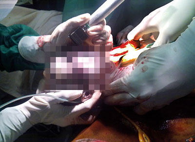 |
| Lump in the neck: Baby's head is delivered while baby is connected to the placenta. |
One such intra-uterine blood transfusion has already been performed by Dr. Dias at a private hospital with Consultant Obstetrician and Gynaecologist Dr.Vijith Vidyabushana in April last year and four more at the De Soysa Hospital for Women with Consultant Obstetricians and Gynaecologists Prof. Hemantha Senanayake and Prof. Athula Kaluarachchi between May and August, MediScene learns.
Recalling one of the cases of a humble mother from Mahiyanganaya whose foetus was detected with anaemia at 25 weeks (six -months plus), he says the need was to keep the pregnancy going till 35 weeks till the baby was born at the De Soysa Hospital. That’s what the intra-uterine blood transfusion did. If foetal anaemia is not treated in the womb the baby will die or survive with severe disabilities.
Stenting – If there is an obstruction to the urine flow of the foetus, the bladder gets distended and the back-pressure of the urine will damage the kidneys. In such cases, a needle is sent through the mother’s tummy into the bladder of the foetus, after which an artificial conduit is made from the bladder to the amniotic cavity (water sac surrounding the foetus) allowing the urine to flow there, releasing the back pressure and saving the kidneys. This procedure can also be performed when the baby’s chest is filled with fluid (pleural effusion).
Twin-to-twin transfusion syndrome – In 20% of all twin pregnancies, the twin foetuses share a single placenta. Known as monochorionic twins, they also share the vascular (blood vessel) connections on the surface of the placenta.
Often the blood flow to both is balanced but in 15% there could be an imbalance with one twin (donor) giving blood to the other (recipient). This leaves the donor hypovolumic and small and the recipient with a fluid overload resulting in heart failure. Both twins are at risk, with 90% of such foetuses dying before 25 weeks (six months plus) if not treated, says Dr. Dias whose doctoral thesis at the University of London is on the ‘Role of ultrasound scanning in twin pregnancy management’.
The answer is to send a fetoscope (a type of telescope) through the mother’s tummy to the womb, identify the community vessels on the placenta surface and block them by Laser.
This procedure through which both twins can be saved in 66% of cases and at least one in 80% is regrettably unavailable in any centre in South Asia, according to Dr. Dias who has brought back with him the tools needed including the fetoscope.
 |
| Baby is delivered completely once the airway is fully secured |
He stresses that the identification or pick-up rate of monochorionic twins is poor as many mothers are not scanned between 11-14 weeks of pregnancy. If the scan is delayed past 14 weeks, there is no way to find out whether it is monochorionic or not, MediScene learns.
Scanning is important when taking into account that 1-2% of all pregnancies are twin pregnancies and with many mothers having babies when they are older or couples seeking in-vitro fertilization (test-tube babies) there is an increase in twin pregnancies.
With an annual total birth rate of 380,000 in Sri Lanka, the formal introduction of scanning between 11 and 14 weeks could save more than 400 monochorionic twins at risk who sometimes do not even get included in the death rates as the perinatal mortality rate is calculated after 28 weeks of pregnancy and the infant mortality rate between birth and one year, it is understood.
When monochorionic twins are identified, monitoring and follow-up action can take place from 16 weeks to prevent the loss of the twins.
Genetic problems – these can be identified in the foetus and categorised as “isolated” or “recurrent” and if recurrent, the parents counselled and advised about future pregnancies. Some cases of this nature were managed together with Clinical Geneticist Prof. Vajira H.W. Dissanayake.
n Foetal medicine also helps to meticulously plan the delivery of the baby so as to avoid or deal with complications.
Citing the case of a foetus with a lump in the neck, Dr. Dias stresses that it is important to plan the delivery as the baby will need immediate resuscitation at birth as the lump may obstruct breathing. If delivered normally, the baby will die, so a multi-disciplinary team will be on hand to provide EX-utero Intrapartum Treatment (EXIT) which is a combination of a Caesarean section delivery and an operation on the baby as he/she is being born.
Referring to such a case handled at the De Soysa Hospital, he explained that doctors from the team of Consultant Paediatrician Dr. R. Ajanthan and Consultant Paediatric Ear, Nose and Throat Surgeon Dr. A.D.K.S.N. Yasawardene were present along with the other medical and nursing staff to help carry out ‘EXIT'. The baby had been diagnosed with an anterior neck lump through a scan earlier and was delivered partially through a Caesarean section. With only the head being taken out of the womb while the baby was still attached to the placenta, the doctors intubated (secured the airway) the baby, after which full delivery took place.
Dr. Dias had been a member of a procedure in the UK where when the intubation did not work the Paediatric Surgeon secured the airway with an immediate tracheostomy, which was later followed by reconstruction surgery.
Importance of ultrasound in foetal care
Ultrasound scanning is essential to monitor the health of the foetus, underscores Dr. Dias, urging that scans between 11-14 weeks should not only be to date the pregnancy but also to manage other aspects until delivery.
All complications such as chromosomal aneuploidy (in 90%), major foetal structural deformities (in 80%), early onset pre-eclampsia (in 90%), foetal growth restrictions (in 70%) and pre-term labour (in 90%), a major killer, can be predicted through scans taken within the first 11-14 weeks of pregnancy, he stresses.
If a foetal heart scan is done between 20-24 weeks, heart disease (in 60%) can be picked up so that the mother can be referred to a tertiary health care centre with all the facilities and a multidisciplinary team including a Consultant Paediatric Cardiologist.
If the baby is delivered at a smaller hospital, serious complications could occur and the baby would die before transfer to such a centre, he says.
Referring to the advancement in the care of expectant mothers, he points out that the ante-natal care model was developed in 1929 because women were dying at childbirth. Most pregnancy complications arose in the latter part of the pregnancy (last trimester or last three months), that is why up to 28 weeks into the pregnancy, the Obstetrician screened the mother; monthly between the 28-36 week period every fortnight and after 36 weeks once a week. Earlier it was assumed that the last trimester complications could not be predicted in the early part of the pregnancy.
But now we know differently, says Dr. Dias who has many research papers to his credit published by prestigious publications including ‘How to date twin pregnancies’ in the ‘British Journal of Obstetrics and Gynaecology’ (BJOG) and ‘How to label twins in the womb’ and ‘Chorionicity’ (determining whether it is mono or dichorionic during the 11-14 week period) in the ‘Ultrasound Obstetrics and Gynaecology’.
The world has moved on, starting the scanning of expectant mothers early, but Sri Lanka seems to be still caught in that outdated protocol, he laments.
The need to implement these vital changes in the Sri Lankan antenatal care protocol was highlighted by Dr. Hemantha Perera, President of the Sri Lanka College of Obstetricians and Gynaecologist at his induction on January 7.
|

