Trigeminal neuralgia: Taking the shooting pain away
Unbearable and excruciating…………it comes in sudden waves, leaving the person grimacing in agony. Just as it comes, it will leave, but the very fear of recurrence is debilitating. The descriptions vary – like a lance hitting the face; sharp and shooting; and as if an electric shock has struck the jaw.
It is Trigeminal neuralgia (TN).
For 20 long years, H.D. Patrick, an electrician, living in Battaramulla had to live with it. From one doctor to another he went but to no avail. “Uhulanna beri, ivasanna beri, amaruvak thibbe. Dath kekkumata wediya daha gunayak wedei,” he says, explaining that it was unbearable and 10 times more than a toothache. The pain was on the right side of his face, above the nose and below the eye. He could not eat, taking blended food and he could hardly talk.

Consultant Neurosurgeon Dr. Jayantha Liyanage
Some doctors gave him tablets and he would carry them in his wallet and pop them every four hours. He also kept the alarm every night and swallowed tablets four-hourly.
Four years ago, two doctors suggested that he see Consultant Neurosurgeon Dr. Jayantha Liyanage who reassured him……“Baya wenna epa. Mama operation ekak karannam.” (Don’t be frightened. I will perform an operation.)
More women than men are hit by this devastating pain, MediScene learns and to find out what this ailment which is murmured about fearfully, we sought out Dr. Liyanage whose skill and technique have helped provide relief to many.
“TN is one of the most or even the ‘most’ agonizing pain a person can suffer,” says Dr. Liyanage, explaining that this “lancinating type of severe pain affects one side of the face”.
Painkillers “hariyanne ne”, is his verdict, as he says that initially, the pain may be trivial or feel like a numbness of the face, progressing to extreme severity, very quickly.
The sufferer, because of the severity, duration and frequency of the pain, feels that life is not worth living, according to Dr. Liyanage. In its wake will follow severe anxiety, affecting even the person’s hygiene, work and life.
In almost all cases the cause is “obscure”, he points out, with the pain generating from the 5th(trigeminal nerve) of the 12 cranial nerves. This is one of the most widely-distributed nerves in the head.
Looking closely at the trigeminal nerve, this Neurosurgeon says that it has three branches that conduct sensations from the upper, middle and lower portions of the face and the oral cavity, to the brain.
The three branches of the trigeminal nerve are:
- Upper or ophthalmic branch – supplying sensation to most of the scalp, forehead and front of the head.
- Middle or maxillary branch – stimulating the cheek, upper jaw, top lip, teeth and gums and also the side of the nose.
- Lower or mandibular branch – supplying nerves to the lower jaw, teeth and gums and bottom lip.

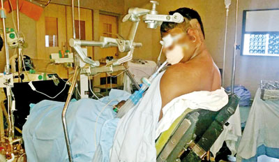
A patient undergoing microvascular decompression. Pix by Sameera Weerasekera
More than one nerve branch can be affected by the disorder, MediScene learns.
Dr. Liyanage says that the contributory factors are unknown, but there is an assumption that it could be due to neuronal damage caused by the vascular loop as the nerve emerges from the brain stem (pons). This could be affecting nerve conduction, propagating severe pain.
The prevalence is about 4 cases per 100,000, in those above 40, with a female preponderance.
“Commonly, the maxillary and mandibular branches of the trigeminal nerve are affected, with the pain pattern being stereotypical (having a sameness) and lasting year after year, making the patient’s life miserable,” he says.
The clinical picture depends on symptoms, as there are no neurological abnormalities. Diagnostic scans, even Magnetic Resonance Imaging (MRI) show nothing, with serological tests being negative. The pain is paroxysmal (on one side) and comes about without any provocation. The ‘triggers’ include simple and routine actions such as talking, brushing of teeth, shaving, eating or drinking cold stuff or even exposure to wind, MediScene understands.
 Dr. Liyanage explains that the clinical presentation is sensitivity in the “trigger-zones” which is a small area of the face, where a light touch can set off the pain. The trigger zones are around the mouth and the chin and just beyond the brow.
The criteria for diagnosis of TN are:
- Paroxysmal attack of pain, lasting seconds to about two minutes, with or without the presence of aching in-between.
- The pain being intense, sharp and stabbing and superficial.
“We take down the history and perform a thorough intraoral examination which includes orthodontic (by referring to a Dental Surgeon) and non-orthodontic examinations,” he says, adding that there is also meticulous examination of the cranial nerves 5, 7 and 8, with the doctors ordering a CT scan and MRI to exclude growths adjacent to the trigeminal nerve.
- Cranial nerve 5 – sensation, corneal reflex and ability to chew will be checked.
- Cranial nerve 7 – musculature of face will be checked by closure of eye, taking mouth to a side and getting the person to blow out air.
Cranial nerve 8 – hearing will be checked.
For Mr. Patrick there is no more pain and there is no carrying of tablets around. Nearly four years ago, on October 23, 2014 he underwent microvascular decompression.
“Devi kenek wage,” is his compliment to Dr. Liyanage for dispelling the pain and the grimacing and paving the way for him to smile again.
| The best option There are two ways of dealing with trigeminal neuralgia, MediScene learns. The two options are pharmacological or surgical.n Pharmacological therapy – Two sets of medications are prescribed, with each set consisting of three medications taken orally. The first set comprises carbamazepine, baclofen and lamotrigine and the second clonazepam, gabapentin and topiramate. Sometimes a cocktail (mix) of these drugs are given.n Surgical option – This has three methods: the minimally-invasive percutaneous stereotactic radiofrequency lesioning (RFL) of the trigeminal ganglion; gamma-knife radiation of the trigeminal route; and microvascular decompression (MVD) through posterior fossa approach. The latter is to relieve abnormal compression of a cranial nerve. Stressing that pharmacological therapy has its limitations due to recurrence, side-effects caused by taking medications long-term and high expense for the patient in buying the drugs, Dr. Jayantha Liyanage points out that the surgical option is better and tolerated well. However, in the surgical option, the gamma-knife radiation is not available in Sri Lanka, while the minimally-invasive RFL does not address the cause, as such with a high chance of pain-recurrence. Dr. Liyange says that MVD, meanwhile, addresses the cause of the pain and brings about a cure. He speaks with evidence in hand, having performed MVD on 15 patients, with even the first patient, H.D. Patrick, who underwent this procedure four years ago having no recurrence of pain. | |
| MVD: Step-by-step explanationConsultant Neurosurgeon Dr. Jayantha Liyanage performs microvascular decompression (MVD), with the patient undergoing the procedure sitting up and not lying down. “Then identification of deep structures is easy, there is less refraction and bleeding is minimal,” he says. Here is a step-by-step explanation from this Neurosurgeon how he performs this operation:
With the exposure of the dura of the brain (the thick membrane that surrounds the brain), the buddy-halo retractor is put into place to keep the axis in the deep structures of the posterior fossa (a small space in the skull, found near the brain stem and cerebellum).
The surgery takes about four hours. |


