A revealing picture
View(s):As we continue our series on common tests with Prof. Shyam Fernando, a Consultant Physician and Professor in Medicine, we shift focus to chest X-rays and what they can reveal. Speaking with MediScene, Prof. Fernando highlights the pros and cons of using this test as a diagnostic tool and explains how it is done as well and what you need to know before you step into the X-ray room.

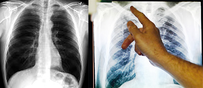
A normal chest X- ray and the X- ray of a tuberculosis patient
What it is:
A chest X-ray is a black-and-white picture of the internal structures of the chest. Unlike a normal photograph, this ‘picture’ is taken using X-rays, a form of radiation, which penetrates the body. Internal structures such as the lungs, heart, blood vessels, lymph nodes, ribs and spine show up on the chest X-ray as shadows. A chest X-ray is a common test done to diagnose diseases of these organs. X-rays are harmless.
How it is it done:
Chest X-ray is done in the radiology department by highly trained technical staff called radiographers. You will be asked to remove necklaces, undress down to the waist and wear a gown. You will have privacy to change clothing. Then you will be asked to stand against a frame and keep still. Usually two pictures are taken; one from the back and the other from a side. You will be alone in the X-ray room, but the radiographer will watch you from behind a screen and give instructions on what to do. Once the ‘picture’ is taken, a specialist doctor called a Radiologist will examine the picture and give a report.
How is it used in diagnosis?
Chest X-ray can detect pneumonia, tumours, tuberculosis, collapsed lung, fluid around the lung and emphysema and ballooned-out lung in long standing smokers. It can also show enlarged heart, fluid around the heart or ballooned out large blood vessels. Narrowed coronary arteries or heart attacks cannot be detected on the chest X-ray. It is also useful to detect fractures of the ribs and the spine.
On the chest X-ray, structures that block radiation appear white, and structures that let radiation through appear black. Bones and the heart will appear white, whereas the lungs filled with air will appear as darker areas. It is not possible for an untrained eye to detect abnormalities. So don’t try to read your own X-ray. You might even get unduly worried when you see some normal white areas, thinking that it is a cancer.
Nowadays, we don’t rely very much on chest X-ray to diagnose heart conditions because we have better tests for that purpose. Your doctor might want to do a chest X-ray to find the cause for chest pain, breathing difficulty, or long standing cough, or when a condition like pneumonia, tuberculosis or lung cancer is suspected. It is also a common test required by the authorities before going for employment abroad. Chest X-rays are also done to assess the progress of a disease condition, its response to treatment and to see whether any complications have arisen.
How do I prepare?
No special preparation is required. It is preferable if you don’t wear any jewellery when you go for the X-ray test. Your doctor would prefer to avoid taking X-rays if there is the slightest chance that you may be pregnant, even though the risk to the baby is negligible. Please inform the radiographer if you might be pregnant or your menstrual period is delayed. A special lead apron will be given to cover the abdomen when taking the X-ray.
What are the after-effects?
It is painless. You don’t feel any sensation as the radiation passes through the body. The risk of exposure to radiation is negligible. There are no after effects.
What are the pitfalls?
A chest X-ray can be completely normal and might fail to reveal diseases (eg. lung cancer) in their early stages if the changes are too small to be detected by the human eye. Even in lung conditions such as asthma, the chest X-ray will be completely normal. It is not a good test to detect heart attacks, heart valve problems or coronary diseases. Your doctor might want to do other tests like ECG, echocardiogram, exercise ECG test, CT scan of the chest, endoscopy or a dye test (series of X-rays done after injecting a dye), such as an angiogram to diagnose your condition.


